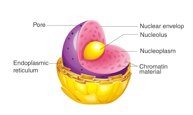
Posts

Nuclear pore

Image
Nuclear pore- ~nuclear pore is a complex structure. ~part of the pore is diaphragm, septum, plug or nucleoplasmin and various annuli. ~annulus is a circular part that covers the nuclear pores. ~a pore with annulus called pore complex. ~sometimes this pore complex is surrounded by a permeable membrane ~ often it is made up of 9 cylinder among which one present at the centre and another 8 present at the peripheral part. ~often this nuclear pore shows network of granular filament ~this pore transfer RNA, ribosome,protein inside and outside the nucleus. Follow our you tube channel #Mogojastro For further topic releted descriptive animated video And also have a unit of sex education https://youtu.be/9igiEz6JDfQ Visit our Facebook page 👇👇👇👇👇👇👇👇👇👇👇👇👇👇👇 https://www.facebook.com/Mogojastro-103461694688339/

Ultrastructure of nucleus

Image
  Ultrastructure of Nucleus 🟦 Introduction A Nucleus is made up of 4 part *Nuclear membrane or karyotheca *Nucleolus *Nuclear sap or nucleoplasm *Chromatin reticulum or nuclear reticulum 🟦 Nuclear membrane or karyotheca *Karyotheca is a bilayered membrane that surrounded nucleus 🟡 structure- 1)made up of lipoprotein 2) this is the trilaminar membrane 3) diameter 62-90 Ã… 4) distance between to memories 100-500 Ã… 5) intermediate space between two membrane is called perinuclear cisternae or perinuclear space. 6) the pores presentation nuclear membrane called nuclear pores 7) diameter of the pores 300-500 Ã… 8) ribosome and endoplasmic reticulum is is attached to it outer membrane of the nuclear membrane 🟡 Nuclear pore- ~nuclear pore is a complex structure. ~part of the pore is diaphragm, septum, plug or nucleoplasmin and various annuli. ~annulus is a circular part that covers the nuclear pores. ~a pore with annulus called pore complex. ~so

Nucleus of cell

Image
NUCLEUS,THE MAJOR PART OF CELL 🟦 Introduction *Nucleus is the biggest organelles of a cell. *It surrounded by a layered plasma membrane  *In the protoplasmic body all hereditary document present. *Nucleus is called the brain of cell. 🟦  Discovery *Leeuwenhoek(first discovered nucleus in RBC of fish blood) *Robert Brown (1831-in the root of orchid,and also named it) *Fontana (1781- discovered nucleolus inside nucleus) *Stasburger(1882-described nucleoplasm) *Flemming (1879- discussed about chromatin of nucleus) *J. Hammerling(1953- discussed about the hereditary influence of nucleus). 🟦 Origin *It is assumed that Nucleus is evolved from protonucleus. *Nuclear membrane is absent in prokaryotic nucleus *only naked chromatin substances (DNA) present in cytoplasm. *In a ideal nucleus chromatin substances place together and surround by nuclear membrane. 🟦 Site *In eukaryotic cells nucleus present at the middle. *but depending upon th

Know what is special in plasma membrane structure

Image
Specialised structure of plasma membrane [A]Microvilli *The cells of epithelium tissue of small intestine, fold Tu forma finger like structure call microvilli *It cell contain 3000 of of that. *Help the small intestine in absorption by increasing its absorption surface area. [Invegination] *This type of invegination all fold found in nephron of kidney *Which type of fold help in absorption of solutes. [Tubules] *Some cell membrane spread into the cytoplasm and former tube like structure called tubule. [Desmosome] *Some part of endoplasmic reticulum store between the intercellular space to form a a tube like structure called desmosome. *Diameter 200m strome *The material which store is called desmosome on macula adherens. *Link between two adjacent cell *Made up of  mucopolysaccharide and protein *Find fibre that is tenofilament or tenofabril came out from desmosome. *Desmosome can find the epidermis layer of skin *when the pressure comes from the outside

Know how fluid mosaic model work

Image
Plasma membrane Fluid mosaic model Synder and Nicolson, two scientist of California university propose the model of plasma membrane in 1972. According to this theory cell membrane is a two layered semi liquid structure why the protein present mosaic. As glacier floats in the ocean, Lipid layer made up of cholesterol,glycolipid, ganglioside. Cell membrane is a jelly like semi liquid structure that made up of phospholipids and there proteins are placed like a mosaic. As a glacier float on ocean cell membrane also float inside the body. This lipid layer has 2 part, *Globular part(hydrophilic in nature) *Longtail part (hydrophobic in nature) *Both the path form a Micelles like structure. As a form of lipid, in the membrane cholesterol, glycolipid, ganglioside present. Protein Protein are of 5 types 1) structural protein 2) channel protein 3) carrier protein 4) receptor protein 5) enzymatic protein Capping of protein The protein of
Image
Cell Membrane   *Nageli and Cramer first discover cell membrane in 1855 *Plowe named the cell membrane in 1931. Origin - The outer layer of cytoplasm converted into cell membrane Appearance - *Observe under electron microscope as trilaminar means it has three layer *Outer and inner part is is electron dense layer or dark layer *Middle part is electron less layer or transparent layer. Structure of cell membrane- *Gorter & Grendel-1926- 2 layerd lipid-protein model  *Danielli(1935) & Schmitt (1940)-3 layerd protein lipid model *Daneielli & Davison (1934,52,70) -2 layed lipid surrounded with 2 layed protein layerd *Robertson(1959) protein-lipid-protein,3 layed unit membrane. Composition *Lipids(20-79%) *Protein (20-70%) *Carbohydrate (1-5%) *water(20%)  Example:RBC Protein-52% Lipids-40% Important component of plasma membrane *Lipids- Phospholipid, sterol, cholesterol, glycolipid spingolipid,etc *Carbohydrates- Branche
Image
Ultrastructure of Cell wall *Main component of cell wall is cellulose *β-D glucose molecule bonded with  β-1,4 glycosidic bond to form cellulose *Is cellulose molecule is 8 A diameter *Michelle form bye joining 100-170 cellulose molecule parallelly *20 Michelle join parallel to form microfibril (250Ã…) *In microwave grill cellulose molecule together form 25 nm finute fibre spindle. *Some microfiber together to form macrofibril *Macrofibrils act as a unit of cell wall *each macrofibrils is 0.4 μm *Each macrofibril contain 5,00,000 cellulose molecule *Under electron microscope it is shown that in cell membrane terminal complex and cellulose synthesis enzyme helps to synthesis cellulose *empty part of macrofiber called matrix *pectin fill the matrix part  by joining a cross link  with hemicellulose. Thickening of cell wall *In secondary cell wall substance like cellulose, lignin, pectin or suberin etc stored to thick cell wall *The substances store